

Abstract
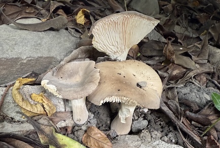
Cantharocybe fudei is introduced as a new species from Taiwan based on both morphological characteristics and phylogenetic analyses of ITS and LSU sequences. It is morphologically similar to C. virosa, with subglobose to ellipsoid basidiospores measuring 8.9–9.3 × 5.7–6 µm but differs in having a flat to depressed pileus, a tapering or club-shaped stipe, and less greyish coloration. Phylogenetic analyses show that it forms a monophyletic lineage sister to the clade of C. virosa and C. habibii. This species is also the first record of the genus Cantharocybe in Taiwan.
References
- Bigelow, H.E. & Smith, A.H. (1973) Cantharocybe, A new genus of Agaricales. Mycologia 65: 485–488. https://doi.org/10.1080/00275514.1973.12019458
- Darriba, D., Posada, D., Kozlov, A.M., Stamatakis, A., Morel, B. & Flouri, T. (2020) ModelTest-NG: A new and scalable tool for the selection of DNA and protein evolutionary models. Molecular Biology and Evolution 37: 291–294. https://doi.org/10.1093/molbev/msz189
- Edler, D., Klein, J., Antonelli, A. & Silvestro, D. (2021) raxmlGUI 2.0: A graphical interface and toolkit for phylogenetic analyses using RAxML. Methods in Ecology and Evolution 12: 373–377. https://doi.org/10.1111/2041-210X.13512
- Ewing, B. & Green, P. (1998) Base-calling of automated sequencer traces using Phred. II. error probabilities. Genome Research 8: 186–194. https://doi.org/10.1101/gr.8.3.186
- Ewing, B., Hillier, L., Wendl, M.C. & Green, P. (1998) Base-calling of automated sequencer traces using Phred. I. accuracy assessment. Genome Research 8: 175–185. https://doi.org/10.1101/gr.8.3.175
- Gardes, M. & Bruns, T.D. (1993) ITS primers with enhanced specificity for basidiomycetes ‐ application to the identification of mycorrhizae and rusts. Molecular Ecology 2: 113–118. https://doi.org/10.1111/j.1365-294x.1993.tb00005.x
- Hosen, M.I., Li, T.H., Lodge, D.J. & Rockefeller, A. (2016) The first ITS phylogeny of the genus Cantharocybe (Agaricales, Hygrophoraceae) with a new record of C. virosa from Bangladesh. MycoKeys 14: 37–50. https://doi.org/10.3897/mycokeys.14.9859
- Hussain, S., Afshan, N.-S., Khan, J., Ahmad, I. & Ahmad, H. (2021) Cantharocybe habibii: a new species in the genus Cantharocybe. Plant Systematics and Evolution 307: 49. https://doi.org/10.1007/s00606-021-01768-x
- Katoh, K., Rozewicki, J. & Yamada, K.D. (2019) MAFFT online service: multiple sequence alignment, interactive sequence choice and visualization. Briefings in Bioinformatics 20: 1160–1166. https://doi.org/10.1093/bib/bbx108
- Kornerup, A. & Wanscher, J.H. (1978) Methuen handbook of colour. Eyre Methuen, London, 252 pp.
- Kozlov, A.M., Darriba, D., Flouri, T., Morel, B. & Stamatakis, A. (2019) RAxML-NG: a fast, scalable and user-friendly tool for maximum likelihood phylogenetic inference. J. Wren (ed.). Bioinformatics 35: 4453–4455. https://doi.org/10.1093/bioinformatics/btz305
- Kumla, J., Suwannarach, N. & Lumyong, S. (2018) Cantharocybe virosa, first record of the genus in Thailand. Mycotaxon 133: 481–485. https://doi.org/10.5248/133.481
- Kuraku, S., Zmasek, C.M., Nishimura, O. & Katoh, K. (2013) aLeaves facilitates on-demand exploration of metazoan gene family trees on MAFFT sequence alignment server with enhanced interactivity. Nucleic Acids Research 41: W22–W28. https://doi.org/10.1093/nar/gkt389
- Lodge, D.J., Padamsee, M., Matheny, P.B., Aime, M.C., Cantrell, S.A., Boertmann, D., Kovalenko, A., Vizzini, A., Dentinger, B.T.M., Kirk, P.M., Ainsworth, A.M., Moncalvo, J.-M., Vilgalys, R., Larsson, E., Lücking, R., Griffith, G.W., Smith, M.E., Norvell, L.L., Desjardin, D.E., Redhead, S.A., Ovrebo, C.L., Lickey, E.B., Ercole, E., Hughes, K.W., Courtecuisse, R., Young, A., Binder, M., Minnis, A.M., Lindner, D.L., Ortiz-Santana, B., Haight, J., Læssøe, T., Baroni, T.J., Geml, J. & Hattori, T. (2014) Molecular phylogeny, morphology, pigment chemistry and ecology in Hygrophoraceae (Agaricales). Fungal Diversity 64: 1–99. https://doi.org/10.1007/s13225-013-0259-0
- Maddison, D.R. & Maddison, W.P. (2021) Chromaseq: a Mesquite package for analyzing sequence chromatograms. Version 1.53. Available from: [http://chromaseq.mesquiteproject.org]
- Maddison, W.P. & Maddison, D.R. (2023) Mesquite: a modular system for evolutionary analysis. Version 3.81. Available from: [https://www.mesquiteproject.org]
- Manimohan, P., Arun Kumar, T.K., Vrinda, K.B. & Pradeep, C.K. (2010) Megacollybia virosa, a new species with toxic basidiomata from India. Mycotaxon 111: 363–368. https://doi.org/10.5248/111.363
- Ovrebo, C.L., Lodge, D.J. & Aime, M.C. (2011) A new Cantharocybe from Belize with notes on the type of Cantharocybe gruberi. Mycologia 103: 1102–1109. https://doi.org/10.3852/10-360
- Parnmen, S., Nooron, N., Leudang, S., Sikaphan, S., Polputpisatkul, D. & Rangsiruji, A. (2020) Phylogenetic evidence revealed Cantharocybe virosa (Agaricales, Hygrophoraceae) as a new clinical record for gastrointestinal mushroom poisoning in Thailand. Toxicological Research 36: 239–248. https://doi.org/10.1007/s43188-019-00024-2
- Ronquist, F., Teslenko, M., Van Der Mark, P., Ayres, D.L., Darling, A., Höhna, S., Larget, B., Liu, L., Suchard, M.A. & Huelsenbeck, J.P. (2012) MrBayes 3.2: efficient Bayesian phylogenetic inference and model choice across a large model space. Systematic Biology 61: 539–542. https://doi.org/10.1093/sysbio/sys029
- Sana, K.M., Mukhtar, F. & Siddique, H. (2025) Scanning electron microscopy and molecular phylogenetic analyses revealed a poisonous mushroom, Cantharocybe virosa, from Pakistan. Microscopy Research and Technique 88: 2763–2770. https://doi.org/10.1002/jemt.70009
- Smith, A.H. (1944) New North American agarics. Mycologia 36: 242–262. https://doi.org/10.1080/00275514.1944.12017545
- Vilgalys, R. & Hester, M. (1990) Rapid genetic identification and mapping of enzymatically amplified ribosomal DNA from several Cryptococcus species. Journal of Bacteriology 172: 4238–4246. https://doi.org/10.1128/jb.172.8.4238-4246.1990
- Vizzini, A., Alvarado, P., Consiglio, G., Marchetti, M. & Xu, J. (2024) Family matters inside the order Agaricales: systematic reorganization and classification of incertae sedis clitocyboid, pleurotoid and tricholomatoid taxa based on an updated 6-gene phylogeny. Studies in Mycology 107: 67–148. https://doi.org/10.3114/sim.2024.107.02
- White, T.J., Bruns, T., Lee, S. & Taylor, J. (1990) Amplification and direct sequencing of fungal ribosomal RNA genes for phylogenetics. In: PCR Protocols. Elsevier, pp. 315–322. https://doi.org/10.1016/B978-0-12-372180-8.50042-1