

Abstract
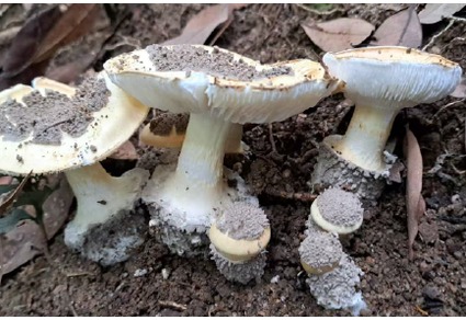
Squamanita hongkongensis is described as a new species from Hong Kong, China, based on morphological evidence and phylogenetic analyses inferred from combined ITS and nrLSU sequences. The new species is morphologically distinguished from all known Squamanita species by the following combination of characters: a cream to pale yellowish pileus bearing grayish-white pyramidal to conical universal veil remnants concentrated at the disc; a nearly smooth, pale yellow stipe; a subglobose mycocecidium (19 × 25 mm), covered with veil remnants similar to those on the pileus, and bearing a membranous, collar-like structure at the stipe-mycocecidium junction; broadly ellipsoid to ellipsoid basidiospores (5.0–6.0 × 3.5–4.7 μm); subfusiform to lageniform pleurocystidia (60–80 × 22–29 μm). Morphological features suggest that the host may belong to Amanita. All phylogenetic analyses recovered S. hongkongensis as sister to Squamanita sp. isolate 2023_42 from North America.
References
- Aime, M.C., Miller, A.N., Aoki, T., Bensch, K., Cai, L., Crous, P.W., Hawksworth, D.L., Hyde, K.D., Kirk, P.M., Lücking, R., May, T.W., Malosso, E., Redhead, S.A., Rossman, A.Y., Stadler, M., Thines, M., Yurkov, A.M., Zhang, N. & Schoch, C.L. (2021) How to publish a new fungal species, or name, version 3.0. IMA Fungus 12: 11. https://doi.org/10.1186/s43008-021-00063-1
- Bas, C. (1965) The genus Squamanita. Persoonia 3: 331–359.
- Bas, C. & Thoen, D. (1998) Squamanita citricolor, a new species from Central Africa. Persoonia 17: 135–139.
- Berkeley, M.J. (1852) Decades of fungi. Decades XXXVII, XXXVIII. Sikkim and Khassya fungi. Hooker’s Journal of Botany and Kew Garden Miscellany 4: 97–107.
- Cui, Y.Y., Cai, Q., Tang, L.P., Liu, J.W. & Yang, Z.L. (2018) The family Amanitaceae: molecular phylogeny, higher-rank taxonomy and the species in China. Fungal Diversity 91: 5–230. https://doi.org/10.1007/s13225-018-0405-9
- Doyle, J.J. & Doyle, J.L. (1987) A rapid DNA isolation procedure for small quantities of fresh leaf tissue. Phytochemical Bulletin 19: 11–15.
- Efron, B. (1979) Bootstrap methods: another look at the jackknife. The Annals of Statistics 7: 1–26. https://doi.org/10.1214/aos/1176344552
- Fayod, V. (1889) Prodrome d’une histoire naturelle des Agaricinés. Annales des Sciences Naturelles, Botanique, sér. 7, 9: 181–411.
- Gardes, M. & Bruns, T.D. (1993) ITS primers with enhanced specificity for basidiomycetes—application to the identification of mycorrhizae and rusts. Molecular Ecology 2: 113–118. https://doi.org/10.1111/j.1365-294X.1993.tb00005.x
- Griffith, G.W., Gajda, K.P., Detheridge, A.P., Douglas, B., Bingham, J., Turner, A., Bowmaker, V., Evans, D.A., McAdoo, W.G. & Dentinger, B.T.M. (2019) Strangler unmasked: parasitism of Cystoderma amianthinum by Squamanita paradoxa and S. pearsonii. Fungal Ecology 39: 131–141. https://doi.org/10.1016/j.funeco.2018.11.012
- Hall, T.A. (1999) BioEdit: a user-friendly biological sequence alignment editor and analysis program for Windows 95/98/NT. Nucleic Acids Symposium Series 41: 95–98.
- Imbach, E.J. (1946) Pilzflora des Kantons Luzern und der angrenzenden Innerschweiz. Mitteilungen der Naturforschenden Gesellschaft Luzern 15: 5–85. https://doi.org/10.5169/seals-523415
- Johnson, M., Zaretskaya, I., Raytselis, Y., Merezhuk, Y., McGinnis, S. & Madden, T.L. (2008) NCBI BLAST: a better web interface. Nucleic Acids Research 36: W5–W9. https://doi.org/10.1093/nar/gkn201
- Katoh, K. & Standley, D.M. (2013) MAFFT multiple sequence alignment software version 7: improvements in performance and usability. Molecular Biology and Evolution 30: 772–780. https://doi.org/10.1093/molbev/mst010
- Kornerup, A. & Wanscher, J.H. (1981) Taschenlexikon der Farben: 1440 Farbnuancen und 600 Farbnamen. Muster-Schmidt, Zürich, 242 pp.
- Kozlov, A.M., Darriba, D., Flouri, T., Morel, B. & Stamatakis, A. (2019) RAxML-NG: a fast, scalable and user-friendly tool for maximum likelihood phylogenetic inference. Bioinformatics 35: 4453–4455. https://doi.org/10.1093/bioinformatics/btz305
- Liu, J.W., Ge, Z.W., Horak, E., Vizzini, A., Halling, R.E., Pan, C.L. & Yang, Z.L. (2021) Squamanitaceae and three new species of Squamanita parasitic on Amanita basidiomes. IMA Fungus 12: 4. https://doi.org/10.1186/s43008-021-00057-z
- Liu, J.W., Luangharn, T., Yu, F.Q. & Bhunjun, C.S. (2024) The complete mitochondrial genome of enigmatic mycoparasitic fungus Squamanita imbachii. Mitochondrial DNA Part B: Resources 9: 716–719. https://doi.org/10.1080/23802359.2024.2356131
- Mondiet, N., Dubois, M.P. & Selosse, M.A. (2007) The enigmatic Squamanita odorata (Agaricales, Basidiomycota) is parasitic on Hebeloma mesophaeum. Mycological Research 111: 599–602. https://doi.org/10.1016/j.mycres.2007.03.009
- Persoon, C.H. (1797) Tentamen dispositionis methodicae fungorum in classes, ordines, genera et familias. Cum supplemento adjecto. P.P. Wolf, Leipzig, 76 pp. https://doi.org/10.5962/bhl.title.42674
- Rathnayaka, A.R., Tennakoon, D.S., Jones, G.E., Wanasinghe, D.N., Bhat, D.J., Priyashantha, A.H., Stephenson, S.L., Tibpromma, S. & Karunarathna, S.C. (2025) Significance of precise documentation of hosts and geospatial data of fungal collections, with an emphasis on plant-associated fungi. New Zealand Journal of Botany 63 (2–3): 462–489. https://doi.org/10.1080/0028825X.2024.2381734
- Redhead, S.A., Ammirati, J.F., Walker, G.R., Norvell, L.L. & Puccio, M.B. (1994) Squamanita contortipes, the Rosetta Stone of a mycoparasitic agaric genus. Canadian Journal of Botany 72: 1812–1824. https://doi.org/10.1139/b94-223
- Ronquist, F., Teslenko, M., van der Mark, P., Ayres, D.L., Darling, A., Höhna, S., Larget, B., Liu, L., Suchard, M.A. & Huelsenbeck, J.P. (2012) MrBayes 3.2: efficient Bayesian phylogenetic inference and model choice across a large model space. Systematic Biology 61: 539–542. https://doi.org/10.1093/sysbio/sys029
- Saar, I., Thorn, R.G., Nagasawa, E., Henkel, T.W. & Cooper, J.A. (2022) A phylogenetic overview of Squamanita, with descriptions of nine new species and four new combinations. Mycologia 114: 769–797. https://doi.org/10.1080/00275514.2022.2059639
- Saccardo, P.A. (1891) Sylloge fungorum omnium hucusque cognitorum. Vol. 9. Supplementum universale, Pars I: Agaricaceae–Laboulbeniaceae. Sumptibus auctoris, Patavii, 1141 pp. https://doi.org/10.5962/bhl.title.5371
- Singer, R. (1973) Diagnoses fungorum novorum Agaricalium III. Beihefte zur Sydowia 7: 1–106.
- Smith, A.H. & Singer, R. (1948) Notes on the genus Cystoderma. Mycologia 40: 454–460. https://doi.org/10.2307/3755152
- Sumstine, D.R. (1914) New or interesting fungi. Mycologia 6: 32–36. https://doi.org/10.1080/00275514.1914.12020946
- Swofford, D.L. (2003) PAUP*. Phylogenetic Analysis Using Parsimony (and Other Methods). Version 4. Sunderland, Massachusetts: Sinauer Associates.
- Tulloss, R.E. (2000) Note sulla metodologia per lo studio del genere Amanita (Agaricales). Bollettino del Gruppo Micologico G. Bresadola, Nuova Serie 43 (2): 41–58.
- Vilgalys, R. & Hester, M. (1990) Rapid genetic identification and mapping of enzymatically amplified ribosomal DNA from several Cryptococcus species. Journal of Bacteriology 172: 4238–4246. https://doi.org/10.1128/jb.172.8.4238-4246.1990
- Wang, H.C. & Yang, Z.L. (2004) Squamanita, a new record to China. Mycosystema 23: 146–148.
- White, T.J., Bruns, T., Lee, S. & Taylor, J. (1990) Amplification and direct sequencing of fungal ribosomal RNA genes for phylogenetics. In: Innis, M.A., Gelfand, D.H., Sninsky, J.J. & White, T.J. (Eds.) PCR protocols: a guide to methods and applications. Academic Press, San Diego, pp. 315–322. https://doi.org/10.1016/B978-0-12-372180-8.50042-1
- Xie, J.M., Chen, Y.R., Cai, G.J., Cai, R.L., Hu, Z. & Wang, H. (2023) Tree visualization by one table (tvBOT): a web application for visualizing, modifying and annotating phylogenetic trees. Nucleic Acids Research 51: W587–W592. https://doi.org/10.1093/nar/gkad359
- Yang, Z.L. (2015) Atlas of the Chinese species of Amanitaceae. Science Press, Beijing, 213 pp.
- Yanisch-Perron, C., Vieira, J. & Messing, J. (1985) Improved M13 phage cloning vectors and host strains: nucleotide sequences of the M13mp18 and pUC19 vectors. Gene 33: 103–119. https://doi.org/10.1016/0378-1119(85)90120-9