
Abstract
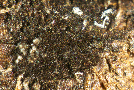
During our ongoing research on fungi associated with sweet cherries in Sichuan Province, China, we collected a saprophytic fungus that was identified as a species of Conioscypha based on ITS region sequence analysis and morphological characteristics. Further phylogenetic analysis based on the nuclear ribosomal internal transcribed spacer (ITS: ITS1-5.8S-ITS2), the nuclear ribosomal small subunit rRNA (SSU), the nuclear ribosomal large subunit rRNA (LSU), the partial translation elongation factor 1-alpha (TEF1), and the partial second largest subunit of RNA polymerase II (RPB2) confirmed that the collection represents a new species within the genus Conioscypha, and herein designated as C. pruni sp. nov. Phylogenetically, Conioscypha pruni forms a clade sister to C. minutispora. Morphologically, C. pruni differs from C. minutispora in having relatively larger conidia with dark-colored oil-drop-like inclusions. The study provided detailed descriptions, photographs of the new species, and an updated phylogeny for the genus Conioscypha.
References
- Blando, F. & Oomah, B.D. (2019) Sweet and sour cherries: Origin, distribution, nutritional composition and health benefits. Trends in Food Science & Technology 86: 517–529. https://doi.org/10.1016/j.tifs.2019.02.052
- Bouckaert, R., Vaughan, T.G., BaridoSottani, J., Duchêne, S., Fourment, M., Gavryushkina, A., Heled, J., Jones, G., Kühnert, D., De Maio, N., Matschiner, M., Mendes, F.K., Müller, N.F., Ogilvie, H.A., du Plessis, L., Popinga, A., Rambaut, A., Rasmussen, D., Siveroni, I., Suchard, M.A., Wu, C.H., Xie, D., Zhang, C., Stadler, T. & Drummond, A.J. (2019) BEAST 2.5: An advanced software platform for Bayesian evolutionary analysis. PLoS Comput Biol 15: e1006650. https://doi.org/10.1371/journal.pcbi.1006650
- CapellaGutiérrez, S., SillaMartínez, J.M. & Gabaldón, T. (2009) trimAl: a tool for automated alignment trimming in large-scale phylogenetic analyses. Bioinformatics 25 (15): 1972–1973. https://doi.org/10.1093/bioinformatics/btp348
- Carbone, I. & Kohn, L. (1999) A method for designing pimer sets for speciation studies in fiamentous ascomycetes. Mycologia 91 (3): 553–556. https://doi.org/10.2307/3761358
- Chen, P., Abeywickrama, P.D., Ji, S., Zhou, Y., Li, X., Zhang, W. & Yan, J. (2023) Molecular identification and pathogenicity of Diaporthe eres and D. hongkongensis (Diaporthales, Ascomycota) associated with cherry trunk diseases in China. Microorganisms 11 (10): 14. https://doi.org/10.3390/microorganisms11102400
- Chuaseeharonnachai, C., Somrithipol, S., Suetrong, S., Klaysuban, A., Pornputtapong, N., Gareth Jones, E.B. & Boonyuen, N. (2017) Conioscypha nakagirii, a new species from naturally submerged wood in Thailand based on morphological and molecular data. Mycoscience 58 (6): 424–431. https://doi.org/10.1016/j.myc.2017.06.003
- Crous, P.W., LuangsaArd, J.J., Wingfield, M.J., Carnegie, A.J., HernándezRestrepo, M., Lombard, L., Roux, J., Barreto, R.W., Baseia, I.G., CanoLira, J.F., Martín, M.P., Morozova, O.V., Stchigel, A.M., Summerell, B.A., Brandrud, T.E., Dima, B., García, D., Giraldo, A., Guarro, J., Gusmão, L.F.P., Khamsuntorn, P., Noordeloos, M.E., Nuankaew, S., Pinruan, U., RodríguezAndrade, E., SouzaMotta, C.M., Thangavel, R., van Iperen, A.L., Abreu, V.P., Accioly, T., Alves, J.L., Andrade, J.P., Bahram, M., Baral, H.O., Barbier, E., Barnes, C.W., Bendiksen, E., Bernard, E., Bezerra, J.D.P., Bezerra, J.L., Bizio, E., Blair, J.E., Bulyonkova, T.M., Cabral, T.S., Caiafa, M.V., Cantillo, T., Colmán, A.A., Conceição, L.B., Cruz, S., Cunha, A.O.B., Darveaux, B.A., da Silva, A.L., da Silva, G.A., da Silva, G.M., da Silva, R.M.F., de Oliveira, R.J.V., Oliveira, R.L., De Souza, J.T., Dueñas, M., Evans, H.C., Epifani, F., Felipe, M.T.C., FernándezLópez, J., Ferreira, B.W., Figueiredo, C.N., Filippova, N.V., Flores, J.A., Gené, J., Ghorbani, G., Gibertoni, T.B., Glushakova, A.M., Healy, R., Huhndorf, S.M., IturrietaGonzález, I., JavanNikkhah, M., Juciano, R.F., Jurjević, Ž., Kachalkin, A.V., Keochanpheng, K., KrisaiGreilhuber, I., Li, Y.C., Lima, A.A., Machado, A.R., Madrid, H., Magalhães, O.M.C., Marbach, P.A.S., Melanda, G.C.S., Miller, A.N., Mongkolsamrit, S., Nascimento, R.P., Oliveira, T.G.L., Ordoñez, M.E., Orzes, R., Palma, M.A., Pearce, C.J., Pereira, O.L., Perrone, G., Peterson, S.W., Pham, T.H.G., Piontelli, E., Pordel, A., Quijada, L., Raja, H.A., Rosas de Paz, E., Ryvarden, L., Saitta, A., Salcedo, S.S., SandovalDenis, M., Santos, T.A.B., Seifert, K.A., Silva, B.D.B., Smith, M.E., Soares, A.M., Sommai, S., Sousa, J.O., Suetrong, S., Susca, A., Tedersoo, L., Telleria, M.T., Thanakitpipattana, D., ValenzuelaLopez, N., Visagie, C.M. & Groenewald, J.Z. (2018) Fungal Planet description sheets: 785–67. Persoonia 41 (108): 238–417. https://doi.org/10.3767/persoonia.2018.41.12
- Cubeta, M.A., Echandi, E., Abernethy, T. & Vilgalys, R. (1991) Characterization of anastomosis groups of binucleate Rhizoctonia species using restriction analysis of an amplified ribosomal RNA gene. Phytopathology 81 (11): 1395–1400. https://doi.org/10.1094/phyto-81-1395
- Egger, K.N. (1995) Molecular analysis of ectomycorrhizal fungal communities. Canadian Journal of Botany 73 (S1): 1415–1422. https://doi.org/10.1139/b95-405
- FAO. (2023) FAO. Available from: https://www.fao.org/faostat/en/#home (accessed 7 August 2023)
- Goh, T.K., Hsieh, S.Y. & Kuo, C.H. (2023) Synonymy of Parafuscosporella with Vanakripa and descriptions of two new species from Taiwan. Mycological Progress 22 (8): 61. https://doi.org/10.1007/s11557-023-01907-3
- Goh, T.K. & Hyde, K.D. (1998) A new hyphomycete genus, Conioscyphopsis, from wood submerged in a freshwater stream and a review of Conioscypha. Mycological Research 102 (3): 308–312. https://doi.org/10.1017/S0953756297004942
- HernándezRestrepo, M., Gené, J., CastañedaRuiz, R., MenaPortales, J., Crous, P. & Guarro, J. (2017) Phylogeny of saprobic microfungi from Southern Europe. Studies in Mycology 86 (1): 53–97. https://doi.org/10.1016/j.simyco.2017.05.002
- Hibbett, D.S. (1996) Phylogenetic evidence for horizontal transmission of group I introns in the nuclear ribosomal DNA of mushroom-forming fungi. Molecular Biology and Evolution 13 (7): 903–17. https://doi.org/10.1093/oxfordjournals.molbev.a025658
- Höhnel, F.X.R. von (1904) Mycologische Fragmente. Mycologische Fragmente 2 (1): 38–60.
- Huelsenbeck, J.P. & Ronquist, F. (2001) MRBAYES: Bayesian inference of phylogenetic trees. Bioinformatics 17 (8): 754–755. https://doi.org/10.1093/bioinformatics/17.8.754
- Hyde, K.D., Dong, Y., Phookamsak, R., Jeewon, R., Bhat, D.J., Jones, E.B.G., Liu, N.G., Abeywickrama, P.D., Mapook, A., Wei, D., Perera, R.H., Manawasinghe, I.S., Pem, D., Bundhun, D., Karunarathna, A., Ekanayaka, A.H., Bao, D.F., Li, J., Samarakoon, M.C., Chaiwan, N., Lin, C.G., Phutthacharoen, K., Zhang, S.N., Senanayake, I.C., Goonasekara, I.D., Thambugala, K.M., Phukhamsakda, C., Tennakoon, D.S., Jiang, H.B., Yang, J., Zeng, M., Huanraluek, N., Liu, J.K., Wijesinghe, S.N., Tian, Q., Tibpromma, S., Brahmanage, R.S., Boonmee, S., Huang, S.K., Thiyagaraja, V., Lu, Y.Z., Jayawardena, R.S., Dong, W., Yang, E.F., Singh, S.K., Singh, S.M., Rana, S., Lad, S.S., Anand, G., Devadatha, B., Niranjan, M., Sarma, V.V., Liimatainen, K., AguirreHudson, B., Niskanen, T., Overall, A., Alvarenga, R.L.M., Gibertoni, T.B., Pfliegler, W.P., Horváth, E., Imre, A., Alves, A.L., da Silva Santos, A.C., Tiago, P.V., Bulgakov, T.S., Wanasinghe, D.N., Bahkali, A.H., Doilom, M., Elgorban, A.M., Maharachchikumbura, S.S.N., Rajeshkumar, K.C., Haelewaters, D., Mortimer, P.E., Zhao, Q., Lumyong, S., Xu, J. & Sheng, J. (2020) Fungal diversity notes 1151–1276: taxonomic and phylogenetic contributions on genera and species of fungal taxa. Fungal Diversity 100 (1): 5–277. https://doi.org/10.1007/s13225-020-00439-5
- Hyde, K.D., Jeewon, R., Chen, Y.J., Bhunjun, C.S., Calabon, M.S., Jiang, H.B., Lin, C.G., Norphanphoun, C., Sysouphanthong, P., Pem, D., Tibpromma, S., Zhang, Q., Doilom, M., Jayawardena, R.S., Liu, J.K., Maharachchikumbura, S.S.N., Phukhamsakda, C., Phookamsak, R., AlSadi, A.M., Thongklang, N., Wang, Y., Gafforov, Y., Gareth Jones, E.B. & Lumyong, S. (2020) The numbers of fungi: is the descriptive curve flattening? Fungal Diversity 103 (1): 219–271. https://doi.org/10.1007/s13225-020-00458-2
- Kalyaanamoorthy, S., Minh, B.Q., Wong, T.K.F., Haeseler, A.V. & Jermiin, L.S. (2017) ModelFinder: fast model selection for accurate phylogenetic estimates. Nature Methods 14 (6): 587–589. https://doi.org/10.1038/nmeth.4285
- Katoh, K. & Standley, D.M. (2013) MAFFT multiple sequence alignment software version 7: improvements in performance and usability. Molecular Biology and Evolution 30 (4): 772–780. https://doi.org/10.1093/molbev/mst010
- Kirk, P.M. (1984) New or interesting microfungi XII. A new species of Conioscypha (Hyphomycetes). Transactions of the British Mycological Society 82 (1): 177–178.
- https://doi.org/10.1016/S0007-1536(84)80230-2
- Li, L., Du, H.Z., Thiyagaraja, V., Bhat, D.J., Phookamsak, R. & Cheewangkoon, R. (2024) Two novel freshwater hyphomycetes, in Acrogenospora (Minutisphaerales, Dothideomycetes) and Conioscypha (Conioscyphales, Sordariomycetes) from Southwestern China. MycoKeys 101 (000): 249–273.
- https://doi.org/10.3897/mycokeys.101.115209
- Liu, N.G., Bhat, D.J., Hyde, K.D. & Liu, J.K. (2019) Conioscypha tenebrosa sp. nov. (Conioscyphaceae) from China and notes on Conioscypha species. Phytotaxa 413 (2): 159–171. https://doi.org/10.11646/phytotaxa.413.2.5
- Liu, N.G., Hyde, K.D., Sun, Y.R., Bhat, D.J., Jones, E.B.G., Jumpathong, J., Lin, C.G., Lu, Y.Z., Yang, J., Liu, L.L., Liu, Z.Y. & Liu, J.K. (2024) Notes, outline, taxonomy and phylogeny of brown-spored hyphomycetes. Fungal Diversity 129 (1): 1–281. https://doi.org/10.1007/s13225-024-00539-6
- Luo, Z.L., Hyde, K.D., Liu, J.K., Maharachchikumbura, S.S.N., Jeewon, R., Bao, D.F., Bhat, D.J., Lin, C.G., Li, W.L., Yang, J., Liu, N.G., Lu, Y.Z., Jayawardena, R.S., Li, J.F. & Su, H.Y. (2019) Freshwater Sordariomycetes. Fungal Diversity 99 (1): 451–660. https://doi.org/10.1007/s13225-019-00438-1
- Matsushima, T. (1996) Matsushima Mycological Memoirs. No. 9. Matsushima Fungus Collection, Kobe, Japan, 120 pp, pl. 8(4)
- Minh, B.Q., Schmidt, H.A., Chernomor, O., Schrempf, D., Woodhams, M.D., von Haeseler, A. & Lanfear, R. (2020) IQ-TREE 2: new models and efficient methods for phylogenetic inference in the genomic era. Molecular Biology and Evolutionl 37 (5): 1530–1534. https://doi.org/10.1093/molbev/msaa015
- Paradis, E., Claude, J. & Strimmer, K. (2004) APE: Analyses of Phylogenetics and Evolution in R language. Bioinformatics 20 (2): 289–290. https://doi.org/10.1093/bioinformatics/btg412
- Pereira, D. & Phillips, A. (2025) Exploring the diversity and ecological dynamics of palm leaf spotting fungi—a case study on ornamental palms in portugal. Journal of Fungi 11 (1): 43. https://doi.org/10.3390/jof11010043
- Réblová, M. & Seifert, K. (2004) Conioscyphascus, a new ascomycetous genus for holomorphs with Conioscypha anamorphs. Studies in Mycology 50 (1): 95–108.
- Réblová, M., Fournier, J. & Štěpánek, V. (2016) Two new lineages of aquatic ascomycetes: Atractospora gen. nov. and Rubellisphaeria gen. et sp. nov., and a sexual morph of Myrmecridium montsegurinum sp. nov. Mycological Progress 15 (3): 21. https://doi.org/10.1007/s11557-016-1166-z
- Rehner, S.A. & Buckley, E. (2005) A Beauveria phylogeny inferred from nuclear ITS and EF1-alpha sequences: evidence for cryptic diversification and links Cordyceps teleomorphs. Mycologia 97 (1): 84–98. https://doi.org/10.3852/mycologia.97.1.84
- Shearer, C.A. (1973) Conioscypha varia Shear. Mycologia 65 (1): 133.
- Shearer, C.A. (1973) Fungi of the Chesapeake Bay and its tributaries II. The genus Conioscypha. Mycologia 65 (1): 128–136. https://doi.org/10.1080/00275514.1973.12019411
- Shearer, C.A. & Motta, J.J. (1973) Ultrastructure and conidiogenesis in Conioscypha (Hyphomycetes). Botany 51 (10): 1747–1751. https://doi.org/10.1139/b73-226
- Srivastava, K.K., Kumar, D. & Barman, P. (2019) Sweet cherry cultivars influencing the growth and productivity under HDP. Journal of Horticultural Sciences 14 (1): 43–47. https://doi.org/10.24154/jhs.v14i1.694
- Subramanian, C.V. (1979) Phialidic Hyphomycetes and Their Teleomorphs – An Analysis. In: Kendrick, B. (Ed.) The Whole Fungus, Vol.1. National Museums of Canada, Ottawa, pp. 125–151.
- Sun, Y.R., Hyde, K.D., Liu, N.G., Jayawardena, R.S., Wijayawardene, N.N., Ma, J., Zhang, Q., AlOtibi, F. & Wang, Y. (2025) Micro-fungi in southern China and northern Thailand: emphasis on medicinal plants. Fungal Diversity 131 (1): 99–299. https://doi.org/10.1007/s13225-024-00549-4
- Toju, H., Tanabe, A.S., Yamamoto, S. & Sato, H. (2012) High-coverage ITS primers for the DNA-based identification of ascomycetes and basidiomycetes in environmental samples. PLoS One 7 (7): e40863. https://doi.org/10.1371/journal.pone.0040863
- Turland, N.J., Wiersema, J.H., Barrie, F.R., Greuter, W., Hawksworth, D.L., Herendeen, P.S., Knapp, S., Kusber, W.H., Li, D.Z., Marhold, K., May, T.W., McNeill, J., Monro, A.M., Prado, J., Price, M.J. & Smith, G.F. (Eds.) (2018) International Code of Nomenclature for algae, fungi, and plants (Shenzhen Code) adopted by the Nineteenth International Botanical Congress Shenzhen, China, July 2017. Regnum Vegetabile 159. Glashütten: Koeltz Botanical Books. https://doi.org/10.12705/Code.2018
- Vilgalys, R. & Hester, M. (1990) Rapid genetic identification and mapping of enzymatically amplified ribosomal DNA from several Cryptococcus species. Journal of Bacteriology 172 (8): 4238–4246. https://doi.org/10.1128/jb.172.8.4238-4246.1990
- Wani, A.A., Singh, P., Gul, K., Wani, M.H. & Langowski, H.C. (2014) Sweet cherry (Prunus avium): Critical factors affecting the composition and shelf life. Food Packaging and Shelf Life 1 (1): 86–99. https://doi.org/10.1016/j.fpsl.2014.01.005
- Xu, R.J., Thiyagaraja, V., Li, Y., Zhou, D.Q., Boonmee, S. & Zhao, Q. (2024) Two novel lignicolous freshwater fungi, Conioscypha xizangensis and Cordana linzhiensis, from the Tibetan Plateau, China. New Zealand Journal of Botany 62 (2–3): 426–442. https://doi.org/10.1080/0028825X.2024.2336044
- Yu, F.M., Jayawardena, R.S., Luangharn, T., Zeng, X.Y., Li, C.J.Y., Bao, S.X., Ba, H., Zhou, D.Q., Tang, S.M., Hyde, K.D. & Zhao, Q. (2024) Species diversity of fungal pathogens on cultivated mushrooms: a case study on morels (Morchella, Pezizales). Fungal Diversity 125 (1): 157–220. https://doi.org/10.1007/s13225-023-00531-6
- Yu, G. (2020) Using ggtree to visualize data on tree-like structures. Current Protocols in Bioinformatics 69: e96. https://doi.org/10.1002/cpbi.96
- Yu, X.D., Sn, Z., Xd, L., Zhu, J.T., Kd, H. & Liu, J.K. (2024) Bambusicolous Fungi from Southwestern China. Mycosphere 15 (1): 5038–5145. https://doi.org/10.5943/mycosphere/15/1/24
- Yu, X.D., Zhang, S.N. & Liu, J.K. (2023) Additions to bambusicolous fungi of Savoryellaceae from Southwest China. Journal of Fungi 9 (5): 571. https://doi.org/10.3390/jof9050571
- Yu, X.D., Zhang, S.N. & Liu, J.K. (2024) Novel hyphomycetous fungi associated with bamboo from Sichuan, China. Phytotaxa 634 (3): 235–254. https://doi.org/10.11646/phytotaxa.634.3.4
- Zelski, S.E., Raja, H.A., Miller, A.N. & Shearer, C.A. (2015) Conioscypha peruviana sp. nov., its phylogenetic placement based on 28S rRNA gene, and a report of Conioscypha gracilis comb. nov. from Peru. Mycoscience 56 (3): 319–325. https://doi.org/10.1016/j.myc.2014.09.002