Abstract
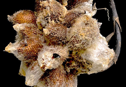
The genus Peniophora is known to be distributed from boreal to tropical regions worldwide. During surveys of plant diseases in southern Thailand in 2022 and 2023, two specimens of rotten snake fruits (Salacca zalacca) were collected. The new species, P. salaccae, is described based on collections with morphological and molecular data. This species is characterized by orange-gray to brownish-orange basidiomes, simple-septate generative hyphae, brown lamprocystidia, subclavate to subcylindrical gloeocystidia, and subtriangular basidiospores. It can be distinguished from P. trigonosperma by its larger basidiospores and shorter lamprocystidia. Molecular phylogenetic analyses of the internal transcribed spacers (ITS) and the large subunit (nrLSU) of the nuclear ribosomal DNA (rDNA) confirmed the position of the new species within the genus Peniophora. The full description, color photographs, illustrations, and the phylogenetic tree showing the position of P. salaccae are provided. Additionally, pathogenicity tests showed that P. salaccae could infect snake fruits, which developed the same symptoms under artificial inoculation conditions as those observed in the field.
References
- Ahmad, R.Z., Setyabudi, D.A. & Wulandari, N.F. (2018) The mold causing agent of rotten snake fruit (Salacca zalacca Gaertn.) from traditional fruit markets. IOP Conference Series: Earth and Environmental Science 197: e012031. https://doi.org/10.1088/1755-1315/197/1/012031
- Andreasen, M. & Hallenberg, N. (2009) A taxonomic survey of the Peniophoraceae. Synopsis Fungorum 26: 56–119.
- Boidin, J. & Lanquetin P. (1983) Two new specis of Peniophora without clamp-connexions. Transactions of the British Mycological Society 81: 279–284. https://doi.org/10.1016/S0007-1536(83)80080-1
- Boidin, J., Mugnier, J. & Canales, R. (1998) Taxonomie molecularies des Aphyllophorales. Mycotaxon 66: 445–491.
- Darriba, D., Taboada, G.L., Doallo, R. & Posada, D. (2012) jModelTest 2: more models, new heuristics and parallel computing. Nature Methods 9: 772. https://doi.org/10.1038/nmeth.2109
- Edgar, R.C. (2004) MUSCLE: multiple sequence alignment with high accuracy and high throughput. Nucleic Acids Research 32: 1792–1797. https://doi.org/10.1093/nar/gkh340
- Felsenstein, J. (1985) Confidence intervals on phylogenetics: an approach using bootstrap. Evolution 39: 783–791. https://doi.org/10.1111/j.1558-5646.1985.tb00420.x
- Grum-Grzhimaylo, O.A., Debets, A.J. & Bilanenko, E.N. (2016) The diversity of microfungi in peatlands originated from the White Sea. Mycologia 108: 233–254. https://doi.org/10.3852/14-346
- Hall, B.G. (2013) Building phylogenetic trees from molecular data with MEGA. Molecular Biology and Evolution 30: 1229–1235. https://doi.org/10.1093/molbev/mst012
- Jeewon, R. & Hyde, K.D. (2016) Establishing species boundaries and new taxa among fungi: Recommendations to resolve taxonomic ambiguities. Mycosphere 7: 1669–1677. https://doi.org/10.5943/mycosphere/7/11/4
- Kornerup, A. & Wanscher, J.H. (1978) Methuen handbook of colour, 3rd ed. Eyre Methuen, London, 252 pp.
- Larsson, E. & Larsson, K.H. (2003) Phylogenetic relationships of russuloid basidiomycetes with emphasis on aphyllophoralean taxa. Mycologia 95: 1037–1065. https://doi.org/10.1080/15572536.2004.11833020
- Larsson, K.H. (2007) Re-thinking the classification of corticioid fungi. Mycological Research 111: 1040–1063. https://doi.org/10.1016/j.mycres.2007.08.001.
- Liu, S.L. & He, S.H. (2018) Taxonomy and phylogeny of Dichostereum (Russulales), with descriptions of three new species from southern China. MycoKeys 40: 111–126. https://doi.org/10.3897/mycokeys.40.28700
- Lygis, V., Vasiliauskas, R., Larsson, K.H. & Stenlid, J. (2005) Wood-inhabiting fungi in stems of Fraxinus excelsior in declining ash stands of northern Lithuania, with particular reference to Armillaria cepistipes. Scandinavian Journal of Forest Research 20: 337–346. https://doi.org/10.1080/02827580510036238
- Miller, S.L., Larsson, E., Larsson, K.H., Verbeken, A. & Nuytinck, J. (2006) Perspectives in the new Russulales. Mycologia 98: 960–970. https://doi.org/10.1080/15572536.2006.11832625
- Rambaut, A. (2012) FigTree. Available from: http://tree.bio.ed.ac.uk/software/figtree/ (accessed 3 June 2024)
- Robles, C.A., Carmarán, C.C. & Lopez, S.E. (2011) Screening of xylophagous fungi associated with Platanus acerifolia in urban landscapes: Biodiversity and potential biodeterioration. Landscape and Urban Planning 100: 129–135. https://doi.org/10.1016/j.landurbplan.2010.12.003
- Ronquist, F., Teslenko, M., Van der Mark, P., Ayres, D.L., Darling, A., Höhna, S., Larget, B., Liu, L., Suchard, M.A. & Huelsenbeck, J.P. (2012) MrBayes 3.2: efficient Bayesian phylogenetic inference and model choice across a large model space. Systematic Biology 61: 539–542. https://doi.org/10.1093/sysbio/sys029
- Safari, Z.S., Ding, P., Nakasha, J.J. & Yuso, S.F. (2020) Combining chitosan and vanillin to retain postharvest quality of tomato fruit during ambient temperature storage. Coatings 10: e1222. https://doi.org/10.3390/coatings10121222
- Spirin, V. & Kout, J. (2015) Duportella lassa sp. nov. from Northeast Asia. Mycotaxon 130: 483–488. https://doi.org/10.5248/130.483
- Stamatakis, A. (2006) RAxML-VI-HPC: maximum likelihood-based phylogenetic analyses with thousands of taxa and mixed models. Bioinformatics 22: 2688–2690. https://doi.org/10.1093/bioinformatics/btl446
- Tamura, K., Stecher, G., Petersen, D., Filipsky, A. & Kumar, S. (2013) MEGA6: molecular evolutionary genetics analysis version 6.0. Molecular Biology and Evolutionary 30: 2725–2729. https://doi.org/10.1093/molbev/mst197
- Vasiliauskas, R., Larsson, E., Larsson, K.H. & Stenlid, J. (2005) Persistence and long-term impact of Rotstop biological control agent on mycodiversity in Picea abies stumps. Biological Control 32: 295–304. https://doi.org/10.1016/j.biocontrol.2004.10.008
- Vilgalys, R. & Hester, M. (1990) Rapid genetic identification and mapping of enzymatically amplified ribosomal DNA from several Cryptococcus species. Journal of Bacteriology 172: 4238–4246. https://doi.org/10.1128/jb.172.8.4238-4246.1990
- Vu, D., Groenewald, M., De Vries, M., Gehrmann, T., Stielow, B., Eberhardt, U., Al-Hatmi, A., Groenewald, J.Z., Cardinali, G., Houbraken, J., Boekhout, T., Crous, P.W., Robert, V. & Verkley, G.J.M. (2019) Large-scale generation and analysis of filamentous fungal DNA barcodes boosts coverage for kingdom fungi and reveals thresholds for fungal species and higher taxon delimitation. Study in Mycology 91: 23–36. https://doi.org/10.1016/j.simyco.2018.05.001
- White, T.J., Burns, T., Lee, S. & Taylor, J.W. (1990) Amplification and direct sequencing of fungal ribosomal RNA genes for phylogenetics. In: Innis, M.A., Gelfand, D.H., Sninsky, J.J. & White, T.J. (Eds.) PCR protocols: a guide to methods and applications. Academic Press, New York, pp. 315–322. https://doi.org/10.1016/B978-0-12-372180-8.50042-1
- Wu, S.H. (2002) A study of Peniophora species with simple-septate hyphae occurring in Taiwan. Mycotaxon 85: 187–199.
- Wulandari, N.F. & Ahmad, R.Z. (2018) Thielaviopsis spp. from Salak [Salacca zalacca (Gaerntn.) Voss] in Indonesia. International Journal of Agricultural Technology 14: 797–804.
- Xu, Y.-L., Tian, Y. & He, S.-H. (2023) Taxonomy and phylogeny of Peniophora Sensu Lato (Russulales, Basidiomycota). Journal of Fungi 9: e93 https://doi.org/10.3390/jof9010093
- Zou, L., Zhang, X., Deng, Y. & Zhao, C. (2022) Four new wood-inhabiting fungal species of Peniophoraceae (Russulales, Basidiomycota) from the Yunnan-Guizhou Plateau, China. Journal of Fungi 8: e1227. https://doi.org/10.3390/jof8111227


