Abstract
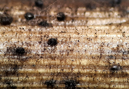
Stemphylium species are saprophytes, or plant pathogens mostly associated with economically important crops. In this study, we isolated Stemphylium strains from grey to brown lesions on the culms of Schoenoplectus sp. (Cyperaceae, Poales). Based on combined morphological characteristics and sequence data of the internal transcribed spacer (ITS‒rDNA) region, parts of glyceraldehyde-3-phosphate dehydrogenase (gapdh) and calmodulin (cmdA) genes, the studied Stemphylium strains were identified and described herein as a new species, S. persianum. The new species is phylogenetically closely related to S. halophilum and S. lycii, however, they are distinguishable based on conidiophores, conidia, and ascospores morphology. Detailed morphological descriptions, illustrations, and phylogenetic relationships with other Stemphylium species are provided herein.
References
- Ahmadpour, A., Ghosta, Y. & Poursafar, A. (2021) Novel species of Alternaria section Nimbya from Iran as revealed by morphological and molecular data. Mycologia 113: 1073–1088. https://doi.org/10.1080/00275514.2021.1923299
- Al-Doory, Y. & Ramsey, S. (1987) Mould and health, who is at risk? Charles C. Thomas Publisher, Springfield, IL.
- Ariyawansa, H.A., Thambugala, K.M., Manamgoda, D.S., Jayawardena, R., Camporesi, E., Boonmee, S., Wanasinghe, D.N., Phookamsak, R., Hongsanan, S., Singtripop, C., Chukeatirote, E., Kang, J.C., Jones, E.B.G. & Hyde, K.D. (2015) Fungal diversity notes 111-252-taxonomic and phylogenetic contributions to fungal taxa. Fungal Diversity 75: 275–277. https://doi.org/10.1007/s13225-015-0323-z
- Berbee, M.L., Pirseyedi, M. & Hubbard, S. (1999) Cochliobolus phylogenetics and the origin of known, highly virulent pathogens, inferred from ITS and glyceraldehyde-3-phosphate dehydrogenase gene sequences. Mycologia 91: 964–977. https://doi.org/10.2307/3761627
- Bessadat, N., Hamon, B., Bataillé-Simoneau, N., Colou, J., Mabrouk, K. & Simoneau, P. (2022) Characterization of Stemphylium spp. associated with tomato foliar diseases in Algeria. Phytopathologia Mediterranea 61: 39–53. https://doi.org/10.36253/phyto-13033
- Brahmanage, R.S., Dayarathne, M.C., Wanasinghe, D.N., Thambugala, K.M., Jeewon, R., Chethana, K.W.T., Samarakoon, M.C., Tennakoon, D.S., De Silva, N.I., Camporesi, E., Raza, M., Yan, J.Y. & Hyde, K.D. (2020) Taxonomic novelties of saprobic Pleosporales from selected dicotyledons and grasses. Mycosphere 11: 2481–2541. https://doi.org/10.5943/mycosphere/11/1/15
- Brahmanage, R.S., Wanasinghe, D.N., Dayarathne, M.C., Jeewon, R., Brahmanage, R.S., Wanasinghe, D.N., Dayarathne, M.C., Jeewon, R., Yan, J., Bulgakov, T.S., Camporesi, E., Kakumyan, P., Hyde, K.D. & Li, X. (2019) Morphology and phylogeny reveal Stemphylium dianthi sp. nov. and new host records for the sexual morphs of S. beticola, S. gracilariae, S. simmonsii and S. vesicarium from Italy and Russia. Phytotaxa 411: 243–263. https://doi.org/10.11646/phytotaxa.411.4.1
- Câmara, M.P.S., O’Neill, N.R. & van Berkum, P. (2002) Phylogeny of Stemphylium spp. based on ITS and glyceraldehyde-3-phosphate dehydrogenase gene sequences. Mycologia 94: 660–672. https://doi.org/10.1080/15572536.2003.11833194
- Chaisrisook, C., Stuteville, D.L. & Skinner, D.Z. (1995) Five Stemphylium spp. pathogenic to alfalfa: occurrence in the United States and time requirements for ascospore production. Plant Disease 79: 369–392. https://doi.org/10.1094/PD-79-0369
- Costa, L.R., Johnson, J.R., Baur, M.E. & Beadle, R.E. (2006) Temporal clinical exacerbation of summer pasture-associated recurrent airway obstruction and relationship with climate and aeroallergens in horses. American Journal of Veterinary Research 67: 1635–1642. https://doi.org/10.2460/ajvr.67.9.1635
- Crous, P.W. & Groenewald, J.Z. (2017) The Genera of Fungi—G 4: Camarosporium and Dothiora. IMA Fungus 8: 131–152. https://doi.org/10.5598/imafungus.2017.08.01.10
- Crous, P.W., Cowan, D.A., Maggs-Kölling, G., Yilmaz, N., Larsson, E., Angelini, C., Brandrud, T.E., Dearnaley, J.D.W., Dima, B., Dovana, F. & Fechner, N. (2020) Fungal Planet description sheets: 1112–1181. Persoonia 45: 1–251. https://doi.org/10.3767/persoonia.2020.45.10
- Crous, P.W., Gams, W., Stalpers, J.A, Robert, V. & Stegehuis, G. (2004) MycoBank: an online initiative to launch mycology into the 21st century. Studies in Mycology 50: 19–22.
- De Gruyter, J., Woudenberg, J.H.C., Aveskamp, M.M., Verkley, G.J.M., Groenewald, J.Z. & Crous, P.W. (2013) Redisposition of phoma-like anamorphs in Pleosporales. Studies in Mycology 75: 1–36. https://doi.org/10.3114/sim0004
- Ershad, D. (2009) Fungi of Iran. 3nd ed. Agricultural Research. Education & Extension Organization, Publication. No. 10, Tehran, 531 pp.
- Farr, D.F., Rossman, A.Y. & Castlebury, L.A. (2024) United States National Fungus Collections Fungus-Host Dataset. Available from: https://nt.ars-grin.gov/fungaldatabases/ (accessed 20 February 2024). [https://fungi.ars.usda.gov/]
- Hay, F., Stricker, S., Gossen, B.D., McDonald, M.R., Heck, D., Hoepting, C., Sharma, S. & Pethybridge, S. (2021) Stemphylium leaf blight: A re-emerging threat to onion production in eastern North America. Plant Disease 105: 3780–3794.
- Hosen, M.I., Ahmed, A.U., Zaman, J., Ghosh, S. & Hossain, K.M.K. (2009) Cultural and physiological variation between isolates of Stemphylium botryosum the causal of Stemphylium blight disease of lentil (Lens culinaris). World Journal of Agricultural Sciences 5: 94–98.
- Inderbitzin, P., Mehta, Y.R. & Berbee, M.L. (2009) Pleospora species with Stemphylium anamorphs: a four-locus phylogeny resolves new lineages yet does not distinguish among species in the Pleospora herbarum clade. Mycologia 101: 329–339. https://doi.org/10.3852/08-071
- Index Fungorum (2024) Available from: http://www.indexfungorum.org/names/names.asp (accessed 20 February 2024)
- Karimzadeh, S. & Fotouhifar, K.B. (2021) Report of some fungi of Pleosporaceae family associated with leaf spot symptoms of plants in Chaharmahal and Bakhtiari province, Iran. Journal of Crop Protection 10: 319–340. https://doi.org/20.1001.1.22519041.2021.10.2.2.8
- Katoh, K., Rozewicki, J. & Yamada, K.D. (2019) MAFFT online service: multiple sequence alignment, interactive sequence choice and visualization. Briefings in Bioinformatics 20: 1160–1166. https://doi.org/10.1093/bib/bbx108
- Lawrence, D.P., Gannibal, P.B., Peever, T.L. & Pryor, B.M. (2013) The sections of Alternaria: formalizing species-group concepts. Mycologia 105: 530–546. https://doi.org/10.3852/12-249
- Leach, C.M. & Aragaki, M. (1970) Effects of temperature on conidium characteristics of Ulocladium chartarum and Stemphylium floridanum. Mycologia 62: 1071–1076.
- Llorente, I. & Montesinos, E. (2006) Brown spot of pear: an emerging disease of economic importance in Europe. Plant Disease 90: 1368–1375. https://doi.org/10.1094/PD-90-1368
- Maddison, W.P. & Maddison, D.R. (2019) Mesquite: a modular system for evolutionary analysis. Version 3.61. [cited 2020 Sep 16]. Available from: http://www.mesquiteproject.org
- Marin-Felix, Y., Hernández-Restrepo, M., Iturrieta-González, I., García, D., Gené, J., Groenewald, J.Z., Cai, L., Chen, Q., Quaedvlieg, W., Schumacher, R.K. & Taylor, P.W.J. (2019) Genera of phytopathogenic fungi: GOPHY 3. Studies in Mycology 94: 1–124. https://doi.org/10.1016/j.simyco.2019.05.001
- Miller, M.A., Pfeiffer, W. & Schwartz, T. (2012) The CIPRES science gateway: enabling high-impact science for phylogenetics researchers with limited resources. Paper presented at: Proceedings of the 1st Conference of the Extreme Science and Engineering Discovery Environment: Bridging from the extreme to the campus and beyond (ACM).
- Misawa, T., Kurose, D., Kubo, C. & Uematsu, S. (2018) First report of Stemphylium herbarum and S. lycopersici causing purple leaf spot of carnation in Japan. New Disease Reports 38: 12. https://doi.org/10.5197/j.2044-0588.2018.038.012
- Moragrega, C., Puig, M., Ruz, L., Montesinos, E. & Llorente, I. (2018) Epidemiological features and trends of brown spot of pear disease based on the diversity of pathogen populations and climate change effects. Phytopathology 108: 223–233. https://doi.org/10.1094/PHYTO-03-17-0079-R
- Nasehi, A., Kadir, J.B., Esfahani, M.N., Mahmodi, F., Ghadirian, H., Ashtiani, F.A. & Soleimani, N. (2013) Leaf spot on lettuce (Lactuca sativa) caused by Stemphylium solani, a new disease in Malaysia. Plant Disease 97: 689. https://doi.org/10.1094/PDIS-10-12-0902-PDN
- Nylander, J.A.A. (2004) Mr Modeltest 2.2: Program distributed by the author. Evolutionary Biology Centre, Uppsala University, Sweden.
- Qian, N., Feng, C., Cheng, Y., Zhang, G., Bhadauria, V., Lu, X. & Zhao, W. (2023) Two species of Stemphylium, S. astragali and S. henanense sp. nov., causing leaf and stem spot disease on Astragalus sinicus in China. Phytopathology 113: 945–952. https://doi.org/10.1094/PHYTO-07-22-0243-SC
- Pei, Y.F., Wang, Y., Geng, Y., O’Neill, N.R. & Zhang, X.G. (2011) Three novel species of Stemphylium from Sinkiang, China: their morphological and molecular characterization. Mycological Progress 10: 163–173. https://doi.org/10.1007/s11557-010-0686-1
- Phukhamsakda, C., McKenzie, E.H., Phillips, A.J., Gareth Jones, E.B., Jayarama Bhat, D., Stadler, M., Bhunjun, C.S., Wanasinghe, D.N., Thongbai, B., Camporesi, E. & Ertz, D. (2020) Microfungi associated with Clematis (Ranunculaceae) with an integrated approach to delimiting species boundaries. Fungal Diversity 102: 1–203. https://doi.org/10.1007/s13225-020-00448-4
- Poursafar, A., Ghosta, Y. & Javan-Nikkhah, M. (2016) A taxonomic study on Stemphylium species associated with black (sooty) head mold of wheat and barley in Iran. Mycologia Iranica 3: 99–109. https://doi.org/ 10.22043/MI.2017.26183
- Poursafar, A., Ghosta, Y. & Javan-Nikkhah, M. (2018) An emended description of Stemphylium amaranthi with its first record for Iran mycobiota. Phytotaxa 371: 93–101. https://doi.org/10.11646/phytotaxa.371.2.3
- Rambaut, A. (2019) FigTree, a graphical viewer of phylogenetic trees. Available from: http://tree.bio.ed.ac.uk/software/figtree (accessed 22 May 2024)
- Rayner, R.W. (1970) A mycological colour chart. Kew, UK: Commonwealth Mycological Institute, 34 pp.
- Ronquist, F., Teslenko, M., Van Der Mark, P., Ayres, D.L., Darling, A., Höhna, S., Larget, B., Liu, L., Suchard, M.A. & Huelsenbeck, J.P. (2012) MrBayes 3.2: efficient Bayesian phylogenetic inference and model choice across a large model space. Systematic Biology 61: 539–542. https://doi.org/10.1093/sysbio/sys029
- Rossman, A.Y., Crous, P.W., Hyde, K.D., Hawksworth, D.L., Aptroot, A., Bezerra, J.L. & Camporesi, E. (2015) Recommended names for pleomorphic genera in Dothideomycetes. IMA Fungus 6: 507–523. https://doi.org/10.5598/imafungus.2015.06.02.14
- Seaney, R.R. (1973) Birdsfoot trefoil. In: Heath, M.E., Metcalfe, D.S. & Barnes, R.F. (Eds.) Forages the science of grassland agriculture. Ames, Iowa, The Iowa State University Press, pp. 177–188.
- Simmons, E.G. (1969) Perfect states of Stemphylium. Mycologia 61: 1–26. https://doi.org/10.1080/00275514.1969.12018697
- Simmons, E.G. (1989) Perfect states of Stemphylium III. Memoirs of the New York Botanical Garden 49: 305–307.
- Simmons, E.G. (2001) Perfect states of Stemphylium—IV. Harvard Papers in Botany 1: 199–208.
- Simmons, E.G. (2004) Novel dematiaceous hyphomycetes. Studies in Mycology 50: 109–118.
- Stamatakis, A. (2014) RAxML version 8: a tool for phylogenetic analysis and post-analysis of large phylogenies. Bioinformatics 30: 1312–1313. https://doi.org/10.1093/bioinformatics/btu033
- Stricker, S.M., Gossen, B.D. & McDonald, M.R. (2021) Risk assessment of secondary metabolites produced by fungi in the genus Stemphylium. Canadian Journal of Microbiology 67: 445–450. https://doi.org/10.1139/cjm-2020-0351
- Sutton, D.A., Fothergill, A.W. & Rinaldi, M.G. (1998) Guide to clinically significant fungi. Williams & Wilkins, 471 pp.
- Swofford, D.L. (2002) PAUP, Phylogenetic analysis using parsimony (and other methods). Version 4.0b10. Sunderland, MA, USA, Sinauer Associates.
- Tamura, K., Stecher, G., Peterson, D., Filipski, A. & Kumar, S. (2013) MEGA6: molecular evolutionary genetics analysis version 6.0. Molecular Biology and Evolution 30: 2725–2729. https://doi.org/10.1093/molbev/mst197
- Vaghefi, N., Thompson, S.M., Kimber, R.B., Thomas, G.J., Kant, P., Barbetti, M.J. & van Leur, J.A. (2020) Multi-locus phylogeny and pathogenicity of Stemphylium species associated with legumes in Australia. Mycological Progress 19: 381–396. https://doi.org/10.1007/s11557-020-01566-8
- Wallroth, F.G. (1833) Flora Cryptogamica Germaniae, pars. post. Nurenberg, 923 pp.
- Wang, C.H., Tsai, Y.C., Tsai, I., Chung, C.L., Lin, Y.C., Hung, T.H., Suwannarach, N., Cheewangkoon, R., Elgorban, A.M. & Ariyawansa, H.A. (2021) Stemphylium leaf blight of welsh onion (Allium fistulosum): an emerging disease in Sanxing, Taiwan. Plant Disease 105: 4121–4131. https://doi.org/10.1094/PDIS-11-20-2329-RE
- Webster, J. (1984) Perfect states as an aid to identification of conidial states. Taxonomy of Fungi Proceedings of the International Symposium on Taxonomy of Fungi, University of Madras, Part 2, 653 pp.
- White, T.J., Bruns, T., Lee, S.J.W.T. & Taylor, J.W. (1990) Amplification and direct sequencing of fungal ribosomal RNA genes for phylogenetics. In: Innis, M.A., Gelfand, D.H., Sninsky, J.J. & White, T.J. (Eds.) PCR protocols: a guide to methods and applications. Academic Press, San Diego, pp. 315–322. https://doi.org/10.1016/B978-0-12-372180-8.50042-1
- Woudenberg, J.H.C., Hanse, B., Van Leeuwen, G.C.M., Groenewald, J.Z. & Crous, P.W. (2017) Stemphylium revisited. Studies in Mycology 87: 77–103. https://doi.org/10.1016/j.simyco.2017.06.001


