
Abstract
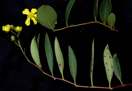
Davilla pygmaea, a new species endemic to the Brazilian Savanna, is described, illustrated and its systematic position is discussed here. The new species is closely related to D. elliptica and D. grandiflora, from which it differs by characters such as a decumbent habit, the shape of its leaf blades, the inflorescence architecture, the size and numbers of flowers, the size of the internal and external sepals during flowering and fruiting, the androecium with 126-131 stamens, and the presence and type of trichomes on vegetative organs. Anatomical characters such as the number of vascular bundles in the petiole and midrib, shape of the midrib in cross section, the margins of the leaf blades, the presence and arrangement of the fibers associated with small-caliber vascular bundles, and the presence of a fiber sheath extension in studied species also differentiate them. Histochemical tests demonstrated that alkaloids, acidic lipids, and phenolic compounds present in the leaf tissues of the new species and D. grandiflora, its closest congener, may have potential medicinal properties. In addition, the new species has its distribution mapped, its flowering and fruiting seasons delimited, and would likely be considered Critically Endangered (CR), as we have found only two populations. A key to identify Davilla species known to Goiás State, Brazil, is provided.
References
Agrios, G.N. (2005) How plants defend themselves against pathogens. In: Agrios, G.N. (Ed.) Plant Pathology, 5th ed. Academic Press, San Diego, pp. 208–248.
Alquini, Y., Bona, C., Boeger, M.R.T., Costa, C.G. & Barra, C.F. (2006) Epiderme. In: Appezzato-da-Glória B, Carmello-Guerreiro SM (eds.) Anatomia vegetal. 2 ed. Ed. UFV, Viçosa, pp. 87–108.
Aymard, G. (1998) Dilleniaceae novae neotropicae VIII. Two new species of Davilla from Brazil. Brittonia 50: 51–55. https://doi. org/10.2307/2807715
Aymard, G. (2002a) Davilla papyracea (Dilleniaceae), a new species from Brazil. Kew Bulletin 57: 487–490. https://doi.org/10.1007/ s12228-008-9046-8
Aymard, G. (2002b) A new species of Davilla (Dilleniaceae) amongst the Flora of SaÞo Paulo, Brazil. Acta Botaìnica Venezuelica 25: 153–159.
Aymard, G. (2007) Three new species of Davilla (Dilleniaceae) from Brazil. Novon 17: 282–287. https://doi.org/10.3417/1055-3177(2007)17[282:TNSODD]2.0.CO;2
Azevedo, A.O., Campos, J.J., Souza, G.G., Veloso, C.C., Duarte, I.D.G., Braga, F.C. & Perez, A.C. (2015) Antinociceptive and anti-inflammatory effects of myricetin 3-O-β-galactoside isolated from Davilla elliptica: involvement of the nitrergic system. Journal of Natural Medicines 69: 487–493. https://doi.org/10.1007/s11418-015-0913-9
Babenko, L.M., Smirnov, O.E., Romanenko, K.O., Trunova, O.K. & Kosakivska, I.V. (2019) Phenolic compounds in Plants: biogenesis and functions. The Ukrainian Biochemical Journal 91: 5–18. https://doi.org/10.15407/ubj91.03.005
Bachman, S., Moat, J., Hill, A.W., de la Torre, J. & Scott, B. (2011) Supporting Red List threat assessments with GeoCAT: geospatial conservation assessment tool. ZooKeys 150: 117–126. https://doi.org/10.3897/zookeys.150.2109
Batalha, M.A. & Mantovani, W. (2001) Floristic composition of the cerrado in the Pé-de-Gigante Reserve (Santa Rita do Passa Quatro, Southeastern Brazil). Acta Botanica Brasilica 15: 289–304
Bieras, A.C. & Sajo, M.G. (2009) Leaf structure of the cerrado (Brazilian savanna) woody plants. Trees 23: 451–471. https:doi.org/10.1007/s00468-008-0295-7
Bobbio, F.O. & Bobbio, P. (2003) Introdução à química de alimentos. 2a edição, Livraria Valeda, São Paulo, 223 pp.
Bukatsch, F. (1972) Bemerkungen zur Doppelfabung Astrablau-Safranin. Microkosmos 61: 255.
Cabané, M., Pireaux, J.-C., Léger, E., Weber, E., Dizengremel, P., Pollet, B. & Lapierre, C. (2004) Condensed lignins are synthesized in poplar leaves exposed to ozone. Plant Physiology 134: 586–594. https: doi.org/10.1104/pp.103.031765
Cain, A.J. (1947) The use of Nile blue in the examination of lipids. The Quarterly Journal of Microscopical Science 88: 383–392. https: doi.org/10.1242/jcs.s3-88.3.383
Chamberlain, C.J. (1932) Methods in plant histology. 5th ed., University of Chicago Press, Chicago, 416 pp. https://doi.org/10.1104/pp.103.031765
Carlos, I.Z., Lopes, F.C.M., Benzatti, F.P., Carli, C.B.A., Marques, M.F., Jordão Junior, C.M., Rinaldo, D., Calvo, T.R., Santos, L.C. & Vilegas, W. (2005) Ação do extrato metanólico e etanólico de Davilla elliptica St.-Hill. (Dilleniaceae) na resposta imune. Brazilian Journal of Pharmacognosy 15: 44–50. https://doi.org/10.1590/S0102-695X2005000100010
Cooley, P.D. (1988) Effects of plant growth rate and leaf lifetime on the amount and type of anti-herbivore defense. Oecologia 74: 531–536. https://doi.org/10.1007/BF00380050
Cronquist, A. (1968) The Evolution and Classification of Flowering Plants. Hoglton Mifflin, Boston, 396 pp.
Davis, K.R. & Hahlbrock, K. (1987) Induction of defense responses in cultured parsley cells by plant cell wall fragments. Plant Physiolgy and Biochemistry 84: 1286–1290. https://doi.org/10.1104/pp.84.4.1286
De Lucia, E.H., Nelson, K., Vogelmann, T.C., Smith, W.K. (1996) Contribution of the intercelular reflectance to photosinthesis in shade leaves. Plant, Cell & Enviroment 19: 159–170. https:doi.org/10.1111/j.1365-3040.1996.tb00237.x
Dickison, W.C. (1969) Comparative morphological studies in Dilleniaceae, IV. Anatomy of node and vascularization of the leaf. Journal of the Arnold Arboretum 50: 384–411.
Dickison, W.C. (1970) Comparative morphological studies in Dilleniaceae, V. Leaf Anatomy. Journal of the Arnold Arboretum 51: 89–101.
Dickison, W.C. (2000) Integrative plant anatomy. Academic Press, San Diego, 533 pp.
Evert, R.F. (2006) Esau’s plant anatomy: meristems, cells and tissues of the plant body: their structure, function and development. 3th ed., Wiley, New Jersey, 601 pp.
Figueiredo, A.C.S., Barroso, J.M.G., Pedro, L.M.G. & Ascensão, L. (2007) Histoquímica e citoquímica em plantas: princípios e protocolos. Lisboa: Faculdade de Ciências da Universidade de Lisboa, Centro de Biotecnologia Vegetal, 68 pp.
Fisher, D.B. (1968) Protein staining of ribboned epon sections for light microscopy. Histochemie 16: 92–96. https:doi.org/10.1007/BF00306214
Fraga, C.N. (2008) Three new species of Davilla (Dilleniaceae) from Bahia, Brazil. Brittonia 60: 355–361. https://doi.org/10.1007/ s12228-008-9046-8
Fraga, C.N. (2012) Filogenia e revisão taxonômica de Davilla Vand. (Dilleniaceae). PhD Thesis, Universidade Federal de Minas Gerais, Belo Horizonte, Brazil. 422 pp.
Fraga, C.N., Aymard, G. & Stehmann, J.R. (2017) Davilla hirsuticarpa (Dilleniaceae), a new species from the Atlantic Forest of Brazil. Plant Ecology and Evolution 150: 367–373. https://doi. org/10.5091/plecevo.2017.1326
Fraga, C.N. & Stehmann, J.R. (2010) Novidades taxonômicas para Dilleniaceae Salisb. Brasileiras. Rodrigueìsia 61: 1–6. https://doi.org/10.1590/2175-7860201061123
Fraga, C.N. & Stehmann, J.R. (2018) Wrongly identified material of Davilla macrocarpa (Dilleniaceae) represents two new species from Brazil. Plant Ecology and Evolution 151: 423–433.
Fraga, C.N. (2020) Davilla In: Flora do Brasil 2020. Jardim Botânico do Rio de Janeiro. Available from: https://floradobrasil2020.jbrj.gov.br/FB7338 (Accessed 1 August 2022).
Goren, A., Ashlock, D. & Tetlow, I.J. (2018) Starch formation inside plastids of higher plants. Protoplasma 255: 1855–1876.
Harbone, J.B. (1993) Introduction to ecological biochemistry, 4th ed. London, Academic Press, 318 pp.
Hectors, K., van Oevelen, S., Guisez, Y., Prinsen, E. & Jansen, M.A. (2012) The phytohormone auxin is a component of the regulatory system that controls UV-mediated accumulation of flavonoids and UV-induced morphogenesis. Physiologia Plantarum 145: 594–603. https://doi.org/10.1111/j.1399-3054.2012.01590.x
Horn, J.W. (2009) Phylogenetics of Dilleniaceae using sequence data from four plastid loci (rbcl, infa, rps4, rpl16 intron). International Journal of Plant Science 170: 794–813.
Irwin, R.E., Cook, D., Richardson, L.L., Manson, J.S. & Gardner, D.R. (2014) Secondary compounds in floral rewards of toxic rangeland plants: impacts on pollinators. Journal of Agricultural and Food Chemistry 62: 7335–7344. https://doi.org/10.1021/jf500521w
IUCN Standards and Petitions Subcommittee (2017) Guidelines for using the IUCN Red List Categories and Criteria. Version 13. Prepared by the Standards and Petitions Subcommittee. Available from: http://www.iucnredlist.org/documents/RedListGuidelines. pdf (Accessed 22 March 2022).
Jácome, R.L.R.P., Oliveira, V.D.C., Oliveira, M.A.T., Mariano, M.C.F. & Oliveira, A.B. (2010) Estudo farmacognóstico comparativo das folhas de Davilla elliptica A. St.-Hil. e D. rugosa Poir., Dilleniaceae. Revista Brasileira de Farmacognosia 20: 390–396.
Jensen, W.A. (1962) Botanical histochemistry, principles and practice. W.H. Freeman, San Francisco, 408 pp.
Johansen, D.A. (1940) Plant microtechnique. McGraw-Hill Book Company Inc., New York, 523 pp.
Kraus, J.E. & Arduin, M. (1997) Manual básico de métodos em morfologia vegetal. EDUR (Editora Universidade Rural), Rio de Janeiro, 198 pp.
Kim, H.U. (2020) Lipid Metabolism in Plants. Plants 9: 1–4. https://doi.org/10.3390/plants9070871
Kubitzki, K. (1971) Doliocarpus, Davilla, und verwandte Gattungen (Dilleniaceae). Mitteilungen der Botanischen Staatssammlung München 9: 1–105.
Kubitzki, K. (1973) Neue und bemerkenswerte Neotropische Dilleniaceen. Mitteilungen der Botanischen Staatssammlung München 9: 707–720.
Kushima, H., Nishijima, C.M., Rodrigues, C.M., Rinaldo, D., Sassá, M.F., Bauab, T.M., Stasi, L.C.D., Carlos, I.Z., Brito, A.R.M.S., Vilegas, W. & Hiruma-Lima, C.A. (2009) Davilla elliptica and Davilla nitida: Gastroprotective, anti-inflammatory immunomodulatory and anti-Helicobacter pylori action. Journal of Ethnopharmacology 123: 430–438. https://doi.org/10.1016/j.jep.2009.03.031
Leegood, R.C. (2008) Roles of the bundle sheath cells in leaves C3 plants. Journal of Experimental Botany 59: 1663–1673.
Linnaeus, C. (1753) Species Plantarum. Impensis Laurentii Salvii, Stockholm, 1200 pp.
Loefling, P. (1758) Iter hispanicum. Lars Salvius, Stockholm, 316 pp.
Mace, M.E. & Howell, C.R. (1974) Histochemistryand identification of condensed tannin percursorin roots of cotton seedlings. Phytopathology 64: 1297–1302.
Martius, C.F.P. von (1838) Herbarium Florae Brasiliensis. Plantae brasilienses exsiccatae, quas denominatas, partim diagnosi aut obsevationibus instructas Botanophilis offert Dr. C. Fr. Ph. de Martius. Flora 21: 49–96.
Matsuura, H. & Fett-Neto, A.G. (2015) Plant Alkaloids: Main Features, Toxicity, and Mechanisms of Action. In: Gopalakrishnakone, P., Carlini, C. and Ligabue-Braun, R. (Eds.) Plant Toxins. Springer, Dordrecht, pp. 1–15. https://doi.org/10.1007/978-94-007-6728-7_2-1
Mendes, F.R., Rodrigues, E., Negri, G., Tabach, R. & Carlini, E.A. (2005) “Cipó-caboclo” (Davilla rugosa Poiret): review, phytochemistry and pharmacological initial data. Jornal Brasileiro de Fitomedicina 3: 100–113.
Mendonça, R.C., Filgueiras, T.S. & Fagg, C.W. (2007) Análise florística da Chapada dos Veadeiros. In: Felfili, J.M., Rezende, A.V., Silva Júnior, M.C. (Orgs.) Biogeografia do bioma Cerrado: vegetação e solos da Chapada dos Veadeiros. Brasília, DF: Ed. UnB, pp. 121–237.
Metcalfe, C.R. & Chalk, L. (1983) Anatomy of Dicotyledons. Oxford, Clarendon Press, V. II, 2nd ed., 279 pp.
Michelin, D.C., Iha, S.M., Rinaldo, D., Sannomiy, A.M., Santos, L.C., Vilegas, W. & Salgado, H.R.N. (2005) Antimicrobial activity of Davilla elliptica St. Hil. (Dilleniaceae). Brazilian Journal of Pharmacognosy 15: 209–211. https://doi.org/10.1590/S0102-695X2005000300008
Morretes, B.L. (1966) Contribuição ao estudo da anatomia das folhas de plantas do cerrado II. Boletim de Botânica da Universidade de São Paulo 305: 209–244.
Morretes, B.L. (1969) Contribuição ao estudo da anatomia das folhas de plantas do cerrado III. Boletim de Botânica da Universidade de São Paulo 331: 7–32.
Naczk, M. & Shahidi, F. (2004) Extraction and analysis of phenolics in food. Journal of Chromatography A 1054: 95–111. https://doi.org/10.1016/j.chroma.2004.08.059
Nimer, E. (1989) Clima. In: IBGE. Geografia do Brasil – Região Centro-Oeste. v. 1. IBGE. Rio de Janeiro. pp. 23–34.
Nishijima, C.M., Delella, F.K., Rodrigues, C.M., Rinaldo, D., Lopes-Ferreira, M.V.A., Rocha, L.R.M., Vilegas, W., Felisbino, S.L. & Hiruma-Lima, C.A. (2015) The anti-inflammatory effects of the methanolic extract and fractions from Davilla elliptica St. Hil. (Dilleniaceae) on Bothrops jararaca envenomation. International Journal of Molecular Sciences 16: 12454–12466. https://doi.org/10.3390/ijms160612454
O’Brien, T.P. & McCully, M.E. (1982) The study of plant structure principles and selected methods. Termarcarphi Pty. Ltda, Melbourne. https://doi.org/10.2307/2259948
Oliveira, L.A. (2000) Anatomia foliar das espécies de Dilleniaceae da Estação Ecológica do Panga (Uberlândia – MG). Master’s Thesis, Universidade Federal de Uberlândia, Uberlândia, Minas Gerais, Brazil. 59 pp.
Poiret, J.L.M. (1812) Encyclopédie méthodique. Botanique, Supplement 2. Chez Plomteux, Liège, 876 pp.
Pyykkô, M. (1966) The leaf anatomy of East Patagonian xerophytic plants. Annales Botanici Fennici 3: 453–622.
Pfister, B. & Zeeman, S. (2016) Formation of starch in plant cells. Cellular and Molecular Life Sciences 73: 2781–2807.
QGIS Development Team (2020) Quantum GIS geographic information system. Open Source Geospatial Foundation Project. Available at: http://www.qgis.org/ (Accessed 20 March 2022)
Raes, J., Rohde, A., Christensen, J.H., van de Peer, Y. & Boerjan, W. (2003) Genome-wide characterization of the lignifications toolbox in Arabidopsis. Plant Physiology 133: 1051–1071. https://doi.org/10.1104/pp.103.026484
Ramos, B.H., Silva, K.L.F., Coimbra, R.R., Chagas, D.B. & Ferreira, W.M. (2015) Anatomy and micromorphometry of Caryocar brasiliense leaves. Rodriguésia 66: 87–94. https://doi.org/10.1590/2175-7860201566106
Rinaldo, D., Silva, M.A., Rodrigues, C.M., Calvo, T.R., Sannomiya, M., Santos, L.C., Kushima, H., Hiruma-Lima, C.A., Brito, A.R.M.S. & Vilegas, W. (2006) Preparative separation of flavonoids from the medicinal plant Davilla elliptica St. Hill. by high-speed counter-current chromatography. Química Nova 29: 947–949. https://doi.org/10.1590/S0100-40422006000500011
Rodrigues, C.M., Rinaldo, D., Sannomiya, M., Santos, L.C., Montoro, P., Piacente, S., Pizza, C. & Vilegas, W. (2008) High-performance liquid chromatographic separation and identification of polyphenolic compounds from the infusion of Davilla elliptica St. Hill. Phytochemical Analysis 19: 17–24. https://doi.org/10.1002/pca.1008
Rolander, D. (1756) Doliocarpus en ort af nytt Genus fran America. Kungliga Svenska vetenskapsakademiens handlingar 17: 256–261.
Rossatto, D.R., Kolb, R.M. & Franco, A.C. (2015) Leaf anatomy is associated with the type of growth form in Neotropical savanna plants. Botany 93: 507–518. https://doi.org/10.1139/cjb-2015-0001.
Saint-Hilaire, A. (1825) Flora Brasiliae Meridionalis: accedunt tabulae delineataea Turpinio aerique incisae. Tomos primus, Paris, Apud A. Belin, Bibliopolam, 480 pp.
Saint-Hilaire, A. & Tulasne, L.R. (1842) Reveu de la Florae du Brésil Meridional. Annales des Sciences Naturelles sér. Botanique 2 (17): 129–143.
Scatena, V.L. & Scremin-Dias, E. (2006) Parênquima, Colênquima e Esclerênquima. In: Appezzato-da-Glória, B. & Carmello-Guerreiro, S.M. (eds.) Anatomia vegetal. 2.ed. Ed. UFV, Viçosa, pp. 109–128.
Silva, C.J.R., Vicentini, T.A., Rosa, S.E.S., Pego, W.F.O. & Vasconcelos, J.M. (2019) Análise morfoanatômica e histoquímica de plantas da espécie Davilla lanosa Braga. South American Journal of Basic Education, Technical and Techinological 6: 277–290.
Soares, M.L., Rezende, M.H., Ferreira, H.D., Figueiredo, A.D.L., Bustamante, K.G.L., Bara, M.T.F. & Paula, J.R. (2005) Caracterização farmacognóstica de folhas de Davilla elliptica St. Hil. (Dilleniaceae). Brazilian Journal of Pharmacognosy 15: 352–360. https://doi.org/10.1590/S0102-695X2005000400017
Sousa, J.N., Mafra, V., Marinho, B.M., Guimarães, V.H.D., Oliveira, L.P., Reis, S.T., Costa, T., Vieira, C.R., Paula, A.M.B., Guimarães, A.L.S. & Santos, S.H.S. (2021) Oral treatment with Davilla elliptica A. St.-Hil. leaves improves liver steatosis and lipid metabolism on a diet-induced obese mice model. Phytomedicine Plus 1: 1–7. https://doi.org/10.1016/j.phyplu.2021.100130
Taiz, L. & Zeiger, E. (2009) Fisiologia vegetal 4.ed. Artmed, Porto Alegre, 719 pp.
Thiers, B. (2022 [continuously updated]) Index herbariorum: a global directory of public herbaria and associated staff. New York Botanical Garden’s virtual herbarium. Published at: http://sweetgum.nybg.org/science/ih/ (Accessed 23 March 2022).
Vandelli, D. (1788) Florae Lusitanicae et brasiliensis specimen et epistolae ab eruditis viris Carolo Linné, Antonio de Haen. Ex Typographia Academico-Regia, Conimbricae, Portugal.
van Fleet, D.S. (1950) The cell forms, and their common substance reactions, in the parenchyma-vascular boundary. Bulletin of the Torrey Botanical Club 77: 340–353.
van Fleet, D.S. (1961) Histochemistry and function of the endodermis. The Botanical Review 27: 165–220
Vahl, M. (1794) Symbolae botanicae. Nicolaus Möller & Son, Copenhagen, 104 pp.
Waller, G.R. & Nowacki, E.K. (1978) Alkaloid biology and metabolism in plants. Phytochemistry 18: 707–708. https://doi.org/10.1016/s0031-9422(00)84313-7
Yeats, T.H. & Rose, J.K. (2013) The formation and function of plant cuticles. Plant Physiolgy 163: 5–20. https://doi.org/10.1104/pp.113.222737
Zhao, H.J. & Zou, Q. (2002) Protective effects of exogenous antioxidants and phenolic compounds on photosynthesis of wheat leaves under high irradiance and oxidative stress. Photosynthetica 40: 523–527. https://doi.org/10.1023/A:1024339716382
Zuccarini, J.G. (1832) Plantarum novarum vel minus cognitarum, quae in horto botanico herbarioque regio Monacensi servantur. Abhandlungen der Mathematisch-Physikalischen Classe der Königlich Beyerischen Akademie der Wissenschaften 1: 287–396.